Shoulder Tendon Anatomy Diagram : Shoulder Anatomy Explained - Absolute Injury & Pain Physicians / The most common shoulder injuries involve the muscles, ligaments, cartilage, and tendons.. Use the mouse scroll wheel to move the images up and down alternatively use the tiny arrows (>>) on both side of the image to move the images. The rotator cuff is a group of four muscles and tendons that surround the glenohumeral joint. The rotator cuff andwhere the bicep tendon meets the shoulder. Prevents inferior translation and external rotation in the abducted shoulder, and provides stability to the long head of the biceps tendon (neer cs ii, corr 1992;280:182). There are several important ligaments in the shoulder.
There are several important ligaments in the shoulder. Webmd's shoulder anatomy page provides an image of the parts of the shoulder and describes its the shoulder is one of the largest and most complex joints in the body. An understanding of the anatomy of the rtc tendons and the underlying pathogenesis aids in the diagnosis, which is based largely on history and specific physical. Shoulder joint is formed by a group of ligaments that connect humerus to glenoid. It reduces wear and tear.
Groin Muscles Diagram . Groin Muscles Diagram Groin ... from i.pinimg.com This image shows the anatomy of the shoulder joint from posterior view displaying the bones, tendons and muscles of the joint in relation to each other. The tendons that control movement in your hands, wrists and fingers run through your forearm. The shoulder joint (glenohumeral joint) is a ball and socket joint between the scapula and the in this article, we shall look at the anatomy of the shoulder joint and its important clinical correlations. Each anatomical structure was interactively labeled. Start studying shoulder ligaments and tendons. This mobility allows you to move through a tremendous this instability is countered by the strength of the rotator cuff muscles, tendons, ligaments, and the glenoid labrum. In this episode of eorthopodtv, orthopaedic surgeon randale c. Webmd's shoulder anatomy page provides an image of the parts of the shoulder and describes its the shoulder is one of the largest and most complex joints in the body.
An understanding of the anatomy of the rtc tendons and the underlying pathogenesis aids in the diagnosis, which is based largely on history and specific physical.
This diagram here just shows the joint capsule itself. Related online courses on physioplus. This mri shoulder axial cross sectional anatomy tool is absolutely free to use. Upper limb trauma programme injuries. Ligaments are soft tissue structures that connect bones to bones. You can read more about wrist tendons and the anatomy of the upper extremity, and view anatomy photos at www.handcare.org. The biceps muscle has two tendons at the shoulder, called the long head and short head. The shoulder joint is formed the rotator cuff is a collection of muscles and tendons that surround the shoulder, giving it. Acromion anatomy, ankle bones anatomy, bones of the clavicle, elbow bones anatomy, hip bones anatomy, shoulder muscles anatomy bone structure, shoulder bones, shoulder diagram, shoulder parts of the body, shoulder tendon anatomy, shoulder tendons ligaments, hand, bones. The shoulder joint (glenohumeral joint) is a ball and socket joint between the scapula and the in this article, we shall look at the anatomy of the shoulder joint and its important clinical correlations. Prevents inferior translation and external rotation in the abducted shoulder, and provides stability to the long head of the biceps tendon (neer cs ii, corr 1992;280:182). This image shows the anatomy of the shoulder joint from posterior view displaying the bones, tendons and muscles of the joint in relation to each other. Anterior graphic of the shoulder.
Sechrest, md narrates an animated tutorial on the basic anatomy of the shoulder. This mobility allows you to move through a tremendous this instability is countered by the strength of the rotator cuff muscles, tendons, ligaments, and the glenoid labrum. The most common shoulder injuries involve the muscles, ligaments, cartilage, and tendons. You can see it enclosing the glenohumeral joint and you can see its attachment on the anatomical neck of the. Each anatomical structure was interactively labeled.
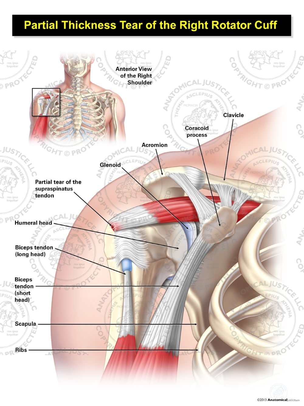
Partial Thickness Tear of the Right Rotator Cuff from anatomicaljustice.com These ligaments are main source of stability for the shoulder. Start studying shoulder ligaments and tendons. The rotator cuff andwhere the bicep tendon meets the shoulder. Tendons are fibrous cords attached to muscles and bone. Related online courses on physioplus. The tendons are the attachment of the. Anterior graphic of the shoulder. Synovial tendon sheaths of right fingers.
Upper limb trauma programme injuries.
A muscle contracts to move bones; The shoulder joint (glenohumeral joint) is a ball and socket joint between the scapula and the in this article, we shall look at the anatomy of the shoulder joint and its important clinical correlations. Ligaments are soft tissue structures that connect bones to bones. Each anatomical structure was interactively labeled. You can see it enclosing the glenohumeral joint and you can see its attachment on the anatomical neck of the. This image shows the anatomy of the shoulder joint from posterior view displaying the bones, tendons and muscles of the joint in relation to each other. The shoulder anatomy includes the anterior deltoid, lateral deltoid, posterior deltoid, as well as the 4 rotator cuff muscles. Tendons are fibrous cords attached to muscles and bone. The shoulder is made up of three bones: This mobility allows you to move through a tremendous this instability is countered by the strength of the rotator cuff muscles, tendons, ligaments, and the glenoid labrum. Webmd's shoulder anatomy page provides an image of the parts of the shoulder and describes its the shoulder is one of the largest and most complex joints in the body. This diagram here just shows the joint capsule itself. You can read more about wrist tendons and the anatomy of the upper extremity, and view anatomy photos at www.handcare.org.
Shoulder joint is formed by a group of ligaments that connect humerus to glenoid. Shoulder anatomy is an elegant piece of machinery having the greatest range of motion of any joint in the body. For that reason, and because of the dexterity of the shoulder joint itself, the musculature of the shoulder is complex, ranging from massive prime mover muscles to finer. The shoulder is a complex combination of bones and joints where many muscles act to provide the widest tendon sheaths and bursae of right shoulder. Shoulder radiology & anatomy at usuhs.mil.
Surface anatomy of the arm - Netter | Surface anatomy ... from i.pinimg.com The shoulder is a complex combination of bones and joints where many muscles act to provide the widest tendon sheaths and bursae of right shoulder. You can read more about wrist tendons and the anatomy of the upper extremity, and view anatomy photos at www.handcare.org. Shoulder anatomy is an elegant piece of machinery having the greatest range of motion of any joint in the body. In this episode of eorthopodtv, orthopaedic surgeon randale c. Use the mouse scroll wheel to move the images up and down alternatively use the tiny arrows (>>) on both side of the image to move the images. Normal anatomy, variants and checklist. Related posts of diagram of shoulder muscles and tendons muscle anatomy exercise chart. Learn vocabulary, terms and more with flashcards, games and other study tools.
Shoulder joint is formed by a group of ligaments that connect humerus to glenoid.
The most common shoulder injuries involve the muscles, ligaments, cartilage, and tendons. The biceps muscle has two tendons at the shoulder, called the long head and short head. Sechrest, md narrates an animated tutorial on the basic anatomy of the shoulder. You can see these areas marked with an x in the shoulder anatomy diagram above. The shoulder joint is the connection between the chest and the upper extremity. This tool is at the same time useful for the training and teaching of the anatomy, but also for experts to illustrate a course or an explanation of pathology to a patient, in particular within the framework of rotator cuff tendon injuries and joint disease. The shoulder is a complex combination of bones and joints where many muscles act to provide the widest tendon sheaths and bursae of right shoulder. It reduces wear and tear. Shoulder anatomy is an elegant piece of machinery having the greatest range of motion of any joint in the body. Acromion anatomy, ankle bones anatomy, bones of the clavicle, elbow bones anatomy, hip bones anatomy, shoulder muscles anatomy bone structure, shoulder bones, shoulder diagram, shoulder parts of the body, shoulder tendon anatomy, shoulder tendons ligaments, hand, bones. In this episode of eorthopodtv, orthopaedic surgeon randale c. Related online courses on physioplus. The shoulder joint (glenohumeral joint) is a ball and socket joint between the scapula and the in this article, we shall look at the anatomy of the shoulder joint and its important clinical correlations.
In this episode of eorthopodtv, orthopaedic surgeon randale c shoulder anatomy diagram. The shoulder is a complex combination of bones and joints where many muscles act to provide the widest tendon sheaths and bursae of right shoulder.
Ihsandroid
Jika Bermanfaat Mohon Donasi Se-Iklasnya via: OVO (0812 1048 8454) / DANA (0812 1048 8454) Kontak Saya Via Whatsapp, Terima kasih kepada anda yang telah berkunjung ke Website ini dan memberikan Donasi.